Our LEGENDplex™ multiplex assays allow you to study the cytokine response associated with the disease. Learn about the principles of the assay and get tips on how to get the best results. Watch now: https://bit.ly/3wc5hHQ


Products

BioLegend's LEGENDplex™ kits are provided as predefined panels, ranging from 3 to 14 specificities, or customers can mix and match any subset within each predefined panel using our Mix and Match system. If you would like to combine specificities across different predefined panels, i.e., Human Cytokine and Human Chemokine, please visit our custom panels tab at the top of this page.

Each kit provides sufficient reagents for at least 100 tests. The assay can be performed in a 96-well filter plate, allowing you to run 40 samples plus 8 standard curve titrations in duplicate, or they can be performed in FACS tubes, where duplicate titrations may not be necessary.

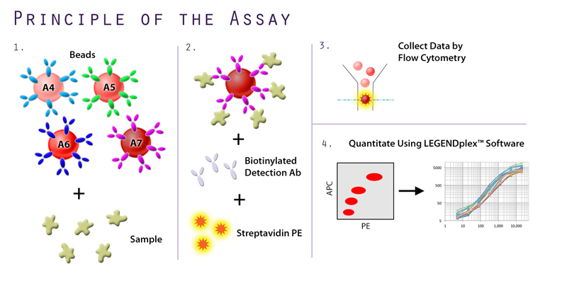
For analysis of LEGENDplex™ assays, BioLegend offers free of charge data analysis software. Please click on the software tab above to learn more about the program options and to request account access.
Principle of the Assay
BioLegend's LEGENDplex™ is a bead-based immunoassay that utilizes the same basic principles of sandwich immunoassays, whereby a soluble analyte is captured between two antibodies. 
  1. Bead populations come in two sizes and differing levels of APC fluorescence, allowing them to be clearly distinguished from each other. Each bead set is conjugated with a specific antibody on the surface and serves as the capture bead for that particular analyte. When a selected panel of capture beads are mixed together and incubated with an unknown sample containing target analytes, each analyte will be bound by its specific capture bead.
  2. After washing, biotinylated detection antibodies are added and each detection antibody will bind to its specific analyte bound on the capture beads, thus forming capture bead-analyte-detection antibody sandwiches.  Streptavidin-phycoerythrin (SA-PE) is subsequently added, which will bind to the biotinylated detection antibodies, providing fluorescent signal with intensities in proportion to the amount of bound analyte.
  3. For each bead population, the PE signal fluorescence intensity is then quantified using a flow cytometer.
  4. The concentration of a particular analyte is determined based on a known standard curve using the LEGENDplex™ data analysis software.