Biolegend

Visualize Neuronal Anatomy
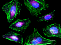
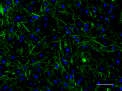
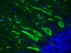
The compartments of neurons can be generally classified into the soma (cell body), axon, dendrite, axonal terminal, and synapse. Reagents for detecting cytoskeletal components like tubulin, actin, and neurofilaments are used to study a neuron’s structural units, or to simply visualize neuronal cells.
Visualization of actin filaments with Flash Phalloidin™ probe
Actin Labeling
Detect actin filaments in neurons with Flash Phalloidin™, an F-actin probe validated for immunofluorescence and immunohistochemistry (IHC). Flash Phalloidin™ is useful for imaging and stabilizing filamentous F-actin in fixed and permeabilized cells.
View Flash Phalloidin™ Data
IHC staining with anti-Neurofilament H & M (NF-H/NF-M), Phosphorylated Antibody
Identify Neurofilament Subunits
The Neurofilament Sampler Kit includes highly validated antibodies for detection of light, medium, heavy, and phosphorylated neurofilament subunits. These Alexa Fluor®-conjugated antibodies can be used for multiplex immunofluorescence (IF) staining to visualize neuronal axons, dendrites, and cell bodies.
Explore the Neurofilament Kit
IHC staining with anti-Tubulin beta 3 (TUBB3) antibody
Gold Standard Microtubule Detection
Tubulin is the main component of microtubules and can be detected with our gold standard antibody clone TUJ1. Tubulin beta 3 (TUBB3) is primarily expressed in neurons in adults, plays an important role in neuronal cell proliferation and differentiation, and is commonly used as a neuronal marker.
Browse TUJ1 conjugates

바이오클론(주)

www.bioclone.co.kr

email : bioclone@bioclone.co.kr

 Flow Cytometry Antibodies is Biolegend !!