Buyer is solely responsible for determining whether Buyer has all intellectual property rights that are necessary for Buyer's intended uses of the BioLegend TotalSeq™ products. For example, for any technology platform Buyer uses with TotalSeq™, it is Buyer's sole responsibility to determine whether it has all necessary third party intellectual property rights to use that platform and TotalSeq™ with that platform.
TotalSeq™-B antibodies are designed to be used with 10x Single Cell 3’ Reagent Kit v3 with Feature Barcoding. The adjusted protocol below is for customers who are utilizing TotalSeq™-A antibodies with the v3 kit instead.
Please read the entire protocol below and the 10x Genomics user guide for the Chromium Single Cell 3ʹ Reagent Kits v3 with Feature Barcoding technology for Cell Surface Protein before starting the experiments. 10x Genomics user guide document number CG000206, Rev D.
Commonly used abbreviations:
- ADT: Antibody derived tags: Antibodies raised against a specific extracellular target which are conjugated to an oligo sequence with the same barcode used to quantify the expression of that target.
- HTO: Hashtag oligonucleotides: A pair of antibodies raised against two different targets which are conjugated to an oligo sequence with the same barcode used to run multiple samples on one 10x lane.
Reagent and Instrument List
For Cell Surface staining:
- TotalSeq™-A antibody-oligo and/or TotalSeq™-A Cell Hashing conjugates
- Biotinylated antibody and oligo barcoded streptavidin
- Human TruStain FcX™ (Fc Receptor Blocking Solution) (BioLegend, Cat# 422301/422302)
- TruStain FcX™ PLUS (anti-mouse CD16/32) (BioLegend, Cat# 156603/156604)
- Phosphate Buffered Saline (PBS) (BioLegend, Cat# 926201 or equivalent)
- Cell Staining Buffer (BioLegend, Cat# 420201)
- 12 x 75mm Falcon™ Round-Bottom Polystyrene Tubes (Fisher Scientific, Cat# 14-959-1A or equivalent)
- Flowmi™ Cell Strainer (Bel-Art, H-B Instrument, Cat# H13680-0040)
For library preparation:
- Quantabio sparQ HiFi PCR Master Mix (2X) (Quantabio, Cat# 95192-250) or KAPA HiFi HotStart ReadyMix (2X) (Kapa Biosystems, Cat# KK2601)
- Quantabio sparQ PureMag Beads (Quantabio, Cat# 951960) or SPRIselect reagent (Beckman Coulter, Cat# B23317)
- 4200 Tapesation (Agilent Technologies, Cat# G2991A)
- DNA High Sensitivity D1000 and High Sensitivity D5000 (Agilent, Cat# 5067-5584/5067-5592)
- Qubit™ 3 (Thermo Fisher Scientific, Cat# Q33226)
- Qubit™ dsDNA HS Assay Kit (Thermo Fisher, Cat# Q32854/Q32851)
- ADT additive primer (0.2µM Stock) and/or HTO additive primer (0.2µM Stock) (See notes at the end of the protocol for further details on primer sequences.)
- TruSeq Small RNA RPIx (10µM Stock) and/or TruSeq D70x_s (10µM Stock) primers (See notes at the end of the protocol for further details on primer sequences.)
Other essential reagents:
- Nuclease-free Water (Thermo Fisher, Cat# AM9937)
- Ethanol (Sigma, Cat# E7023-500ML)
- Nuclease-Free Pipette Tips (e.g. Thermo Fisher Scientific AM12650, AM12660 or equivalent)
- TempAssure PCR 8-strips (USA Scientific, Cat# 1402-4700)
- PCR Thermocycler (Bio-Rad, T100™ Thermal Cycler)
- Countess™ II FL Automated Cell Counter (ThermoFisher, Cat# AMQAF1000)
Researchers are advised to validate equivalent products when substituting for the above recommendations.
Protocol
I) Cell labeling
- Prepare single cell suspension following a suitable protocol
- Resuspend cells at a concentration of 2x10⁷ cells/mL in staining buffer
- Make note of cell viability (>95%) and also include dead cells in the total cell count.
- If high cell death is observed, live cell enrichment (e.g. by Flow Cytometry) is recommended.
- Aliquot 50 µl of cells into 12 x 75mm tubes.
- Add 5 µl of Human TruStain FcX™ Fc, or 0.5 µl of TruStain FcX™ PLUS (anti-mouse CD16/32) Blocking reagent
Note: We no longer recommend the use of dextran during blocking/staining. If you have any questions please contact BioLegend Technical Support.
- Incubate for 10 min at 4°C.
- While cells are incubating in Fc Block, prepare antibody pool using 1 µg (or titrated amounts) of each TotalSeq™ or Biotinylated antibody.
- To maximize performance, centrifuge the antibody pool at 14,000xg at 2 – 8°C for 10 minutes before adding to the cells.
Note: If antibody cocktail volume is less than 50 µl, add Cell Staining Buffer up to 50 µl, then centrifuge.
- Carefully pipette out the liquid, avoiding the bottom of the tube, and add the TotalSeq™ antibody cocktail to the cell suspension.
- Incubate for 30 minutes at 4°C.
- Add 3.5 mL of cell staining buffer and spin 5 minutes 400g at 4°C. Repeat wash 2 more times for a total of 3 washes.
- If using biotinylated antibodies, incubate with the appropriate oligo barcoded streptavidin at the recommended amount specified in the product technical datasheet for 20 minutes. Then add 3.5mLs of cell staining buffer and spin 5 minutes 400g at 4°C. Repeat wash 2 more times for a total of 3 washes.
- Resuspend cells in 500 µL of PBS to get a concentration of 1x10⁶ cells/mL.
- Filter cells through 40 µm Flowmi™ Cell Strainer.
- Verify cell concentration by counting on hemocytometer after filtration.
Note: We highly recommend determining cell viability. Ideally the viability should be >90% for optimal capture rate. The presence of a large number of non-viable cells can decrease the efficiency of the cell partitioning and recovery within the 10x Chromium chip.
- Dilute cells as necessary for appropriate input into the 10X Chromium chip.
II) Run 10x Genomics single cell 3' v3 assay as described through Post Gem-RT Cleanup – Dynabeads (step 2.1). 10x Genomics Document CG000185, Rev D.
At cDNA amplification step (Step 2.2), use the following table:
| |
ADT 1 rxn (µl) |
HTO 1rxn (µl) |
ADT + HTO 1 rxn (µl) |
| Amp Mix |
50 |
50 |
50 |
| cDNA Primers* |
15 |
15 |
15 |
| ADT Additive Primer (0.2 µM stock) |
1 |
0 |
1 |
| HTO Additive primer (0.2 uM stock) |
0 |
1 |
1 |
| Total |
66 |
66 |
67 |
* included with 10x Genomics 3’ kit, different from Feature cDNA primers 2.
Notes:
Follow steps 2.3A and 2.3B exactly to separate ADTs/HTOs from cDNA. Continue to use 70 µL of sparQ or SPRI beads in step 2.3B.
ADT: Antibody derived tags, it refers to regular TotalSeq™-A antibodies
HTO: TotalSeq™-A Hashtag antibodies
See notes at the end of the protocol for further details on primer sequences.
III) ADT and mRNA library preparation
- A) Prepare Sample Index PCR Mix.
| For ADT |
For HTO |
Volume (µl) |
| Purified ADT/HTO fraction |
Purified ADT/HTO fraction |
5 |
SI PCR primer
(10uM stock) |
SI PCR primer
(10uM stock) |
2.5 |
| TrueSeq Small RNA RPIx (10uM stock) |
TruSeq D70x_s
(10uM stock) |
2.5 |
| 2X QuantaBio or Kapa Hifi Master Mix |
2X QuantaBio or Kapa Hifi Master Mix |
50 |
| RNAse-free water |
RNAse-free Water |
40 |
| |
Total |
100 |
Note: For samples that contain both ADT and HTO, perform two separate reactions and add 5 µl of “purified ADT/HTO fraction” from the same sample to ADT or HTO reaction.
-
- B) Incubate in a thermal cycler with the following protocol:
ADT
| 98°C |
2min |
|
| 98°C |
20sec |
14 - 15 Cycles |
| 60°C |
30sec |
| 72°C |
20sec |
| 72°C |
5min |
|
| 4°C |
hold |
|
|
HTO
| 98°C |
2min |
|
| 98°C |
20sec |
12 - 13 Cycles |
| 64°C |
30sec |
| 72°C |
20sec |
| 72°C |
5min |
|
| 4°C |
hold |
|
|
-
- C) Post ADT/HTO library amplification clean-up
- Add 120 µl sparQ or SPRIselect Reagent (1.2X) to each sample.
- Incubate 5 min at room temperature.
- Place on the magnet in its High position until the solution clears.
- Carefully remove and discard the supernatant.
- Place tubes on magnet in its High position. Wash pellet twice with 200 µl 80% ethanol.
- Centrifuge briefly. Place on the magnet Low. Remove remaining ethanol.
- Remove from the magnet. Add 40.5 µl water.
- Incubate 2 min at room temperature.
- Place on the magnet in its Low position until the solution clears.
- Transfer 40 µl to a new tube strip. Store at 4°C for up to 72 h or at −20°C for long-term storage.
- ADT/HTO libraries are now ready to be sequenced
Quantify libraries by standard methods (QuBit, BioAnalyzer, qPCR).
ADT libraries will be around 180 bp (Figure 1B).
Sequencing CITE-seq libraries:
We estimate that an average of 100 molecules per ADT per cell is sufficient to achieve useful information. The number of reads required to obtain 100 molecules depends on the complexity of the sequencing library (e.g. duplication rate). ADT and mRNA cDNA sequencing libraries can be pooled at desired proportions. To obtain sufficient read coverage for both libraries, we typically sequence ADT libraries in 5-10% of a lane and cDNA library fraction at 90% of a lane (HiSeq2500 Rapid Run Mode Flowcell) (Approximately 5k reads/cell for ADTs).
Notes:
Oligonucleotide sequences:
TotalSeq™ antibodies. Each clone is barcoded with a unique oligonucleotide sequence. These contain standard small TruSeq RNA read 2 sequences and can be amplified using Illumina’s Truseq Small RNA primer sets (RPIx – primers, see example RPI1 below)
CCTTGGCACCCGAGAATTCCAAACAAGACCCTTGAGBAAAAAAAAAAAAAAAAAAAAAAAAAAAAAA*A*A.

Please visit https://www.biolegend.com/totalseq for detailed information:
Oligos required for ADT library amplification:
PAGE purification is the preferred method when ordering primers.
The phosphorothioate bonds in the primer renders the oligonucleotide resistant to nuclease degradation.
A unique Illumina primer (index) should be used for each 10x Genomics sample lane used within one sequencing lane.
- 10x Genomics SI-PCR primer 5’AATGATACGGCGACCACCGAGATCTACACTCTTTCCCTACACGACGC*T*C
- ADT cDNA PCR additive primer 5’CCTTGGCACCCGAGAATT*C*C
- HTO cDNA PCR additive primer 5’GTGACTGGAGTTCAGACGTGTGC*T*C
- Illumina Small RNA RPI1 primer (for ADT amplification; i7 index 1, Oligonucleotide sequences, Illumina) 5’CAAGCAGAAGACGGCATACGAGATCGTGATGTGACTGGAGTTCCTTGGCACCCGAGAATTC*C*A
- Illumina TruSeq D701_s primer (for HTO amplification; i7 index 1, shorter than the original Illumina sequence) 5’CAAGCAGAAGACGGCATACGAGATCGAGTAATGTGACTGGAGTTCAGACGTGT*G*C
* indicates a phosphorothioate bond
B indicates C or G or T; not A nucleotide
|
Name
|
Sequence
|
Do Not Use with the
10x Genomics Index
|
|
SI-PCR primer
|
AATGATACGGCGACCACCGAGATCTACACTCTTTCCCTACACGACGC*T*C |
|
|
HTO additive primer
|
GTGACTGGAGTTCAGACGTGTGC*T*C |
|
|
ADT additive primer
|
CCTTGGCACCCGAGAATT*C*C |
|
| |
|
D701_S
|
CAAGCAGAAGACGGCATACGAGATCGAGTAATGTGACTGGAGTTCAGACGTGT*G*C |
|
|
D702_S
|
CAAGCAGAAGACGGCATACGAGATTCTCCGGAGTGACTGGAGTTCAGACGTGT*G*C |
|
|
D703_S
|
CAAGCAGAAGACGGCATACGAGATAATGAGCGGTGACTGGAGTTCAGACGTGT*G*C |
|
|
D704_S
|
CAAGCAGAAGACGGCATACGAGATGGAATCTCGTGACTGGAGTTCAGACGTGT*G*C |
|
|
D705_S
|
CAAGCAGAAGACGGCATACGAGATTTCTGAATGTGACTGGAGTTCAGACGTGT*G*C |
|
|
D706_S
|
CAAGCAGAAGACGGCATACGAGATACGAATTCGTGACTGGAGTTCAGACGTGT*G*C |
|
|
D707_S
|
CAAGCAGAAGACGGCATACGAGATAGCTTCAGGTGACTGGAGTTCAGACGTGT*G*C |
|
|
D708_S
|
CAAGCAGAAGACGGCATACGAGATGCGCATTAGTGACTGGAGTTCAGACGTGT*G*C |
|
|
D709_S
|
CAAGCAGAAGACGGCATACGAGATCATAGCCGGTGACTGGAGTTCAGACGTGT*G*C |
|
|
D710_S
|
CAAGCAGAAGACGGCATACGAGATTTCGCGGAGTGACTGGAGTTCAGACGTGT*G*C |
|
|
D711_S
|
CAAGCAGAAGACGGCATACGAGATGCGCGAGAGTGACTGGAGTTCAGACGTGT*G*C |
SI-GA-E7
|
|
D712_S
|
CAAGCAGAAGACGGCATACGAGATCTATCGCTGTGACTGGAGTTCAGACGTGT*G*C |
SI-GA-A10
|
| |
|
RPI1
|
CAAGCAGAAGACGGCATACGAGATCGTGATGTGACTGGAGTTCCTTGGCACCCGAGAATTC*C*A |
|
|
RPI2
|
CAAGCAGAAGACGGCATACGAGATACATCGGTGACTGGAGTTCCTTGGCACCCGAGAATTC*C*A |
|
|
RPI3
|
CAAGCAGAAGACGGCATACGAGATGCCTAAGTGACTGGAGTTCCTTGGCACCCGAGAATTC*C*A |
|
|
RPI4
|
CAAGCAGAAGACGGCATACGAGATTGGTCAGTGACTGGAGTTCCTTGGCACCCGAGAATTC*C*A |
|
|
RPI5
|
CAAGCAGAAGACGGCATACGAGATCACTGTGTGACTGGAGTTCCTTGGCACCCGAGAATTC*C*A |
|
|
RPI6
|
CAAGCAGAAGACGGCATACGAGATATTGGCGTGACTGGAGTTCCTTGGCACCCGAGAATTC*C*A |
|
|
RPI7
|
CAAGCAGAAGACGGCATACGAGATGATCTGGTGACTGGAGTTCCTTGGCACCCGAGAATTC*C*A |
SI-GA-A11 |
|
RPI8
|
CAAGCAGAAGACGGCATACGAGATTCAAGTGTGACTGGAGTTCCTTGGCACCCGAGAATTC*C*A |
|
Representative Data and Troubleshooting
A.
 |
B.
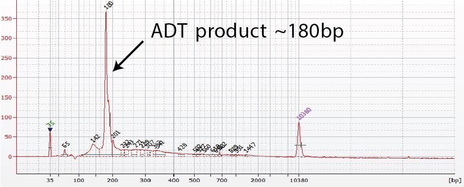 |
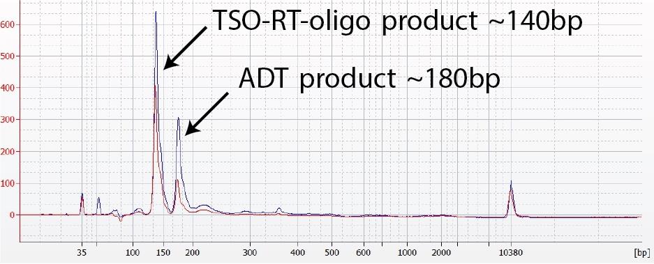
Figure 1. ADT library verification.
(Left graph) A TSO-RT-oligo product (~140 bp) can be amplified during the ADT PCR by carryover primers from cDNA amplification. The product will not cluster but will interfere with quantification. Sequential 2X sparQ or SPRI purification of the ADT fraction after cDNA amplification reduces carryover of primers from cDNA amplification, and minimizes the amplification of this product during ADT-library amplification. To further enrich for ADT specific product the purified ADT library can be reamplified for 3 additional cycles with ADT specific primer sets or P5/P7 generic primers. (Right graph) A clean ADT library will contain a predominant single peak at around 180 bp.
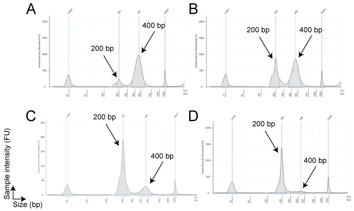
Figure 2. My ADT/HTO library contains a large peak at ~400bp
Overamplification of a library can lead to depletion of available primers and/or dNTPs resulting in self-priming of PCR products by their P5 and/or P7 adapters. This can lead to the production of “daisy-chains” or “bubble products”. These products consist of essentially 2 ADT or HTO barcode sequences attached to one another in 1 long oligo tag that is twice as long as the original oligo tag; these products appear as peaks at approximately 400 bp. These peaks can be more or less pronounced (panel A and B respectively).
The larger peak is perfectly acceptable to sequence. However, it is difficult to quantify these libraries to titrate for sequencing. This error can be corrected by performing another PCR reaction using generic P5/P7 primers (not used in the protocol) for one or two cycles on the 1.2x sparQ or SPRI cleaned up product. The PCR reaction will convert the larger product to the desired size product partially (panel C) or completely (panel D) as the major peak.